Flow Cytometry

Our flow cytometry capability offers analysis and sorting of fluorescently labelled cells, small particles and gentle sorting of large objects and small organisms.
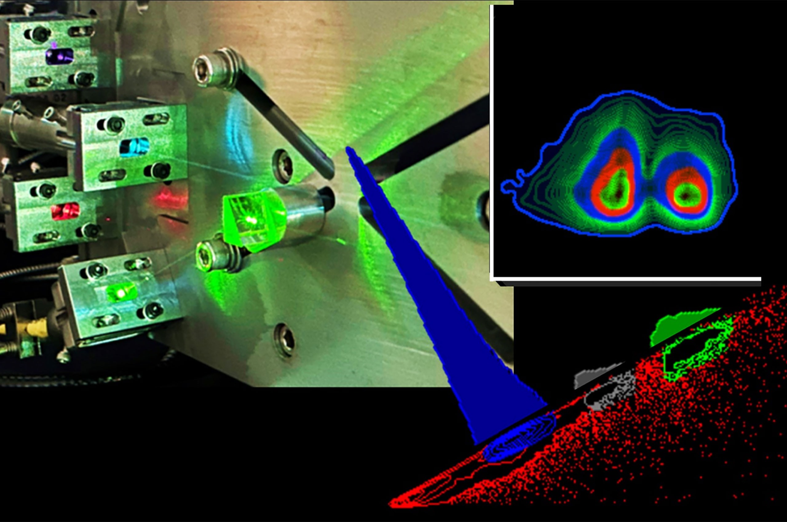
The cytometers measure relative cell size, complexity and fluorescence across multiple channels. Data corresponding from each cell is plotted in histograms and dot plots to enable cell subsets or particles to be precisely defined, compared and quantified.
We work with students, academic researchers and industry partners across all disciplines, including agricultural, pharmaceutical, biomedical and environmental research. Our staff have extensive experience to provide comprehensive training and technical support to help you self-operate our equipment. We also offer end-to-end services from experimental design and sample preparation to data acquisition and data analysis.
We are committed to providing high-quality research support and services to our customers. As part of our ongoing commitment to quality, the facility is currently seeking ISO9001 certification. Read our quality statement.
We can offer:
- cytometry equipment, in a PC2 certified laboratory
- cell sorting and data analysis services
- initial and on-going project consultation
- advice on experimental design and fluorescent panel design
- method development
- FlowJo portal licenses for post-acquisition analysis
- data analysis workshops and training.
- Immunophenotyping
Identify immune cell phenotypes present in complex biological samples in inflammatory disease.
- Cytokine quantitation
Use bead array assays to identify and measure intracellular cytokines in isolated cell populations, or in cell culture supernatants and serum samples to study inflammation and disease.
- Cell cycle analysis
Quantitate and analyse cell cycle status in cancer cells, primary cells or cell lines and plant nuclei. Evaluate the effect of potential drug candidates on cell cycle status.
- Estimate genome size and ploidy
Using isolated nuclei assess genome size and ploidy status of plant tissue, including seeds, leaves and root samples.
- Cell sorting and index sorting
Isolate single populations of cells or nuclei for genomic analysis, PCR or biological assays.
- Detection and analysis of Extracellular Vesicles (EVs)
Detect, and measure the size and surface markers of EVs, exosomes and small particles.
- Apoptosis assays
Detect and quantify apoptotic and necrotic cells.
- Viability assays
Measure and quantify live and dead cells or bacteria.
- Sorting large cells and small organisms
Sort large cells or clusters and small organisms (40 µm-750 µm) including Drosophila larvae into plates or tubes for assay analysis. Sort objects on the basis of size or fluorescent colours. - Detection and quantitation of intracellular phosphoproteins and transcription factors.
FACSCanto II (3-laser)
Lasers: 3 (blue 488nm, violet 405nm and red 633nm)
Detectors: 8 Fluorescent Channels
Sample from Tubes or High Throughput Sampler (HTS) for 96well plates
The laser configuration can be viewed by clicking here.

FACSCanto II (2-laser)
Lasers: 2 (blue 488nm and red 633nm)
Detectors: 5 Fluorescent Channels
Sample from Tubes or High Throughput Sampler (HTS) for 96well plates
The laser configuration can be viewed by clicking here.

CytoFLEX S
2 equipments available
Lasers: 4 (blue 488nm, red 633nm, violet 405nm, yellow/green 561nm)
Detectors: 12 Fluorescent Channels
Sample from Tubes or High Throughput Sampler (HTS) for 96well plates
The laser configuration can be viewed by clicking here.

FACSymphony A3
Lasers: 5 (blue 488nm, red 637nm, violet 405nm, UV 349nm, yellow/green 561nm)
Detectors: 28 Fluorescent Channels
Sample from Tubes or High Throughput Sampler (HTS) for 96well plates
The laser configuration can be viewed by clicking here.

FACSymphony A1
Lasers: 4 (blue 488nm, red 637nm, violet 405nm, yellow/green 561nm)
Detectors: 16 Fluorescent Channels
Small particle detector for extracellular vesicles
Sample from Tubes or High Throughput Sampler (HTS) for 96 well plates
The laser configuration can be viewed by clicking here.

BioSorter
Lasers: 2 (blue 488nm, yellow/green 561nm)
Detectors: 3 Fluorescent Channels
Equipped with 2 (FOCA) Fluidics and Optics Core Assemblies, 500 µm (objects 20-400 µm) and 1000 µm (objects 30-750 µm)
Suitable for sorting small organisms and large cells
Collection tubes: 15 ml and 50 mL
Collection Plates: 24, 48, 96 and 384 well
Collect 1 population

FACSAria III
Lasers: 4 (blue 488nm, red 633nm, violet 405nm, yellow/green 561nm)
Detectors: 14 Fluorescent Channels
Collection tubes: 5, 15 ml and 1.5 mL Eppendorf tubes
Plates: 6,12,24, 96 and 384 well
Slides
Collect up to 4 populations
Nozzles: 70 µm, 85 µm, 100 µm
Cooling system for sample and collected sample
The laser configuration can be viewed by clicking here.

FACSAria Fusion
Contained in a Class II Biological safety cabinet
Lasers: 5 (blue 488nm, red 633nm, violet 405nm, yellow/green 561nm)
Detectors: 17 Fluorescent Channels
Small particle detector for extracellular vesicles
Collection tubes: 5, 15 ml and 1.5 mL Eppendorf tubes
Plates: 6,12, 24, 96 and 384 well
Slides
Collect up to 4 populations
Nozzles: 70 µm, 85 µm,100 µm, 130 µm
Cooling system for sample and collected samples
The laser configuration can be viewed by clicking here.

The Bioimaging Flow Cytometry Facility has contributed to research outputs including publications. Our recent publications include:
- Augusto, D.G., Murdolo, L.D., Chatzileontiadou, D.S.M. et al. A common allele of HLA is associated with asymptomatic SARS-CoV-2 infection. Nature (2023), 620, 128–136. https://doi.org/10.1038/s41586-023-06331-x
- Tran , V., Brettle, H., Diep, H. et al. Sex‑specific effects of a high fat diet on aortic inflammation and dysfunction. Nature Scientific Reports (2023), 13:21644. https://doi.org/10.1038/s41598-023-47903-1
- Day, Z.I., Mayfosh, A. J., Baxter, A.A. et al. Defining a Water-Soluble Formulation of Arachidonic Acid as a Novel Ferroptosis Inducer in Cancer Cells. Biomolecules (2024),14, 555. https://doi.org/10.3390/biom14050555
The Bioimaging Platform also offers expertise and equipment in various imaging capabilities, including:
We have also partnered with the Olivia Newton-John Cancer Research Institute (ONJCRI) which has complementary flow cytometry capabilities that can be accessed through their Flow Cytometry Core Facility.
The platform’s contributions to research outputs (e.g., publications, presentations, posters) should be acknowledged where possible. These contributions could include:
- paid technical help and services
- accessing research equipment
- scientific advice
- writing assistance.
Proper acknowledgement enables us to demonstrate our value to the research community and highlight our impact on research excellence, which is critical to securing continued funding for our services. Our staff are also researchers with extensive experience and citing them helps to advance their careers.
In cases where substantial intellectual and experimental contributions were made by platform staff, co-authorship must also be offered in accordance with the Australian Code for the Responsible Conduct of Research, regardless of whether payment was made for the services. Researchers should also notify the platform of any publications arising from the support provided by our staff, regardless of whether a co-authorship is offered.
Learn more about how to acknowledge us:
All publications resulting from the use of our services and facilities should include this acknowledgement:
‘The authors acknowledge the La Trobe University [Platform Name] for [support received].’
e.g., The authors acknowledge the La Trobe University Proteomics and Metabolomics Platform for the provision of instrumentation, training and technical support.
OR
e.g., The authors acknowledge the La Trobe University Statistics Consultancy Platform for providing advice on statistical analysis.
If you received significant assistance, guidance or help from our platform staff, or where staff have personally generated research data, they should be acknowledged by name:
‘The authors thank [Staff Name] from the La Trobe University [Platform Name] for [his/her/their] support and guidance in this work.’
e.g., The authors thank [Staff Name] from the La Trobe University Proteomics and Metabolomics Platform for collecting and analysing data for proteomics studies, shown in Figure X.
If a platform staff contribute more than just routine techniques or advice, they should be invited to be a co-author on the publications that describe the data. This applies to the development or adaptation of protocols to suit specific experiments, samples or materials, (re)design of experiments, and extensive data analysis and interpretation.
Co-authorship is independent of whether payment was made for the work/ service.
Access
We work with both academic researchers and industry partners, including pharmaceutical and biotechnology companies. We provide a range of access models to suit different needs, including:
- Instrument access
We provide training to users to self-operate our equipment. Users are charged at an hourly rate based on the equipment they use.
- Fee-for-service
We conduct service work for academic and industry researchers, including experimental design (including fluorescence panel advice), sample preparation, data acquisition and analysis. We can tailor our service provision to suit your needs, contact us to discuss your project.
We offer competitive pricing to all researchers. To enquire about our pricing or arrange a different access method, contact us via bioimaging@latrobe.edu.au today.
Contact
For more information about this capability, please contact:
Dr Margaret Veale
Flow Cytometry Specialist
T: +61 3 9479 1733
E: M.Veale@latrobe.edu.au or bioimaging@latrobe.edu.au
