Light Microscopy
The light microscopy facility offers researchers access to a range of high performance confocal and widefield fluorescence microscopes to visualise and study the structure and function of biological samples (e.g., cells, organisms) with high resolution and detail.
We work with academic and industry researchers across all disciplines. We provide expert advice on project planning and experimental design, offer equipment training and support users with basic and advanced microscopy techniques and digital image analysis. We also conduct contracted research for industry partners.
We are committed to providing high-quality research support and services to our customers. As part of our ongoing commitment to quality, the facility is currently seeking ISO9001 certification. Read our quality statement.
We can offer:
- equipment training and access to the facility.
- advice on project planning and experimental design
- initial and on-going project consultation
- method development and validation
- sample preparation service
- data acquisition, digital image analysis, and automated script writing service
- Brightfield and fluorescence slide scanning service
- support in basic and advanced microscopy techniques, including FRET and FRAP
- Super-resolution imaging (<200nm) using Airyscan and SRRF Stream
- Live cell imaging
Real-time monitoring of the dynamic of biological processes in living cells with high-resolution lenses and an incubation system with CO2 and temperature control.
- Multichannel imaging
Study stained cells, 3D cultures, tissues and small organisms using multichannel fluorescence microscopy.
- Interaction and mobility studies
Quantitative analysis of interactions and mobility of molecules in cells using advanced techniques such as FLIM, FRET, FRAP and FCS.
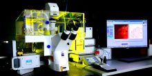
Zeiss LSM 980 upright spectral laser scanning confocal
The Zeiss 980 has two sensitive detectors and four lasers (405, 488, 563, 633 nm) for sample excitation and imaging. It also features a 32-channel spectral detector, as well as a choice of eight high-resolution objectives (5 -100x), including three dipping lenses.
Use it for multichannel fluorescence in animals (e.g. Zebrafish, drosophila) or plants, Z stack 3-D reconstructions and extended focus images, and for creation of tiled mosaic "overview" images. Employ spectral imaging and unmixing to discriminate multiple overlapping fluorescence emission profiles at once.
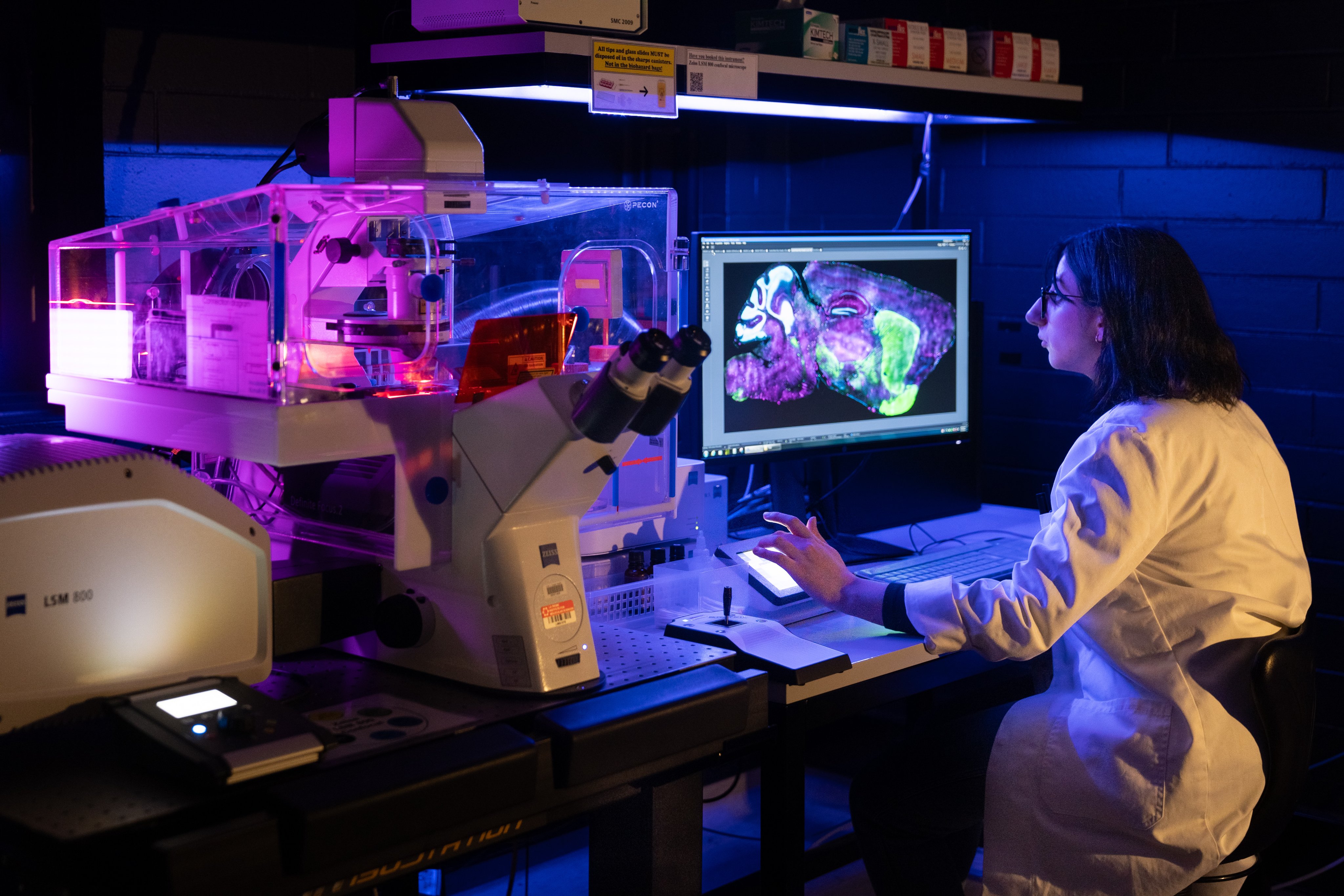
Zeiss LSM 900 airyscan laser scanning confocal
This inverted instrument is equipped with four lasers (405, 488, 563, 633 nm), six high-resolution objective lenses (10x, 20x, 25x multi immersion, 40x water, and 40x and 63x oil), sensitive detectors, a Definite Focus auto-focus module and an incubation system with CO2 and temperature control. The 900 is used for multi-channel fluorescence imaging of fixed tissues, as well as stable long-term live-cell imaging work. Enhanced resolution below the diffraction limit (to about 140 nm) is possible in SR mode by utilising the 32 channel Airyscan detector (1.7-fold resolution improvement = 120 nm).
A high resolution monochrome camera fitted to this microscope also facilitates rapid widefield imaging and navigation of large areas of interest.
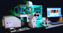
Andor dragonfly 202 spinning disk confocal
The Andor Dragonfly 202 delivers outstanding multi-dimensional images from the subcellular (nm) to whole organism (cm). Coupled with dual Andor Sona sCMOS cameras its unrivalled combination of speed and sensitivity allows researchers to image dynamic events at high temporal resolutions and image live organisms for days.
Integrated super-resolution (<100nm) solutions via SRRF-Stream+ with confocal and widefield are possible with the press of a button. Furthermore, it incorporates the newly developed High Power Laser Engine (HLE) with 7 laser lines and Borealis illumination to allow more even illumination from UV-Vis to NIR and thus more usable data across the field of view. An integrated OKO live cell chamber and Nikon TiE2 inverted microscope with eight available objective lenses and perfect focus system allows for long term, high-resolution imaging of cells and animals.
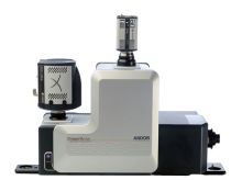
Zeiss LSM 780 laser scanning confocal
The Zeiss 780 inverted microscope has seven laser lines for sample excitation and features three photo multiplier tubes, a 32-channel spectral detector, a choice of five high-resolution objectives (10-100x) and live cell incubator.
Use it for multichannel fluorescence imaging of cells (live or fixed), 3-D reconstructions and extended focus images for imaging of 3-D cultures via confocal Z stacks, and tiling for creation of mosaic "overview" images of larger samples. Employ spectral imaging and unmixing to discriminate multiple overlapping fluorescence emission profiles at once.
Zeiss cell observer spinning disk confocal
The Cell Observer is a hybrid instrument with five laser lines for sample excitation, 20x and 63x objectives, Yokogawa CSU-X1 spinning disk, dual Photometrics EMCCD cameras, and integrated Definite Focus system and a live cell incubator for long-term experiments.
Designed for speed, the spinning disk employs multi-point rather than single-point scanning to illuminate the sample. This allows for 4D imaging experiments where 3-dimensional Z stacks can be captured at every time point (rather than a single focal plane) to gain greater information. Simultaneous 2-channel imaging is possible using synchronised cameras and 3-channel fluorescence imaging with DIC imaging sequentially in one camera mode.
Zeiss Axioscan 7 slide scanner
Our Zeiss Axioscan 7 slide scanner combines high-speed digitisation and outstanding image quality, and a variety of imaging modes in a fully automated and easy to operate system.
Fast batch scanning of up to 100 slides in a single run at magnifications 5x, 10x, 20x, and 40x. Modalities available are colour brightfield, polarization, DIC, phase contrast and seven-channel fluorescence from UV to NIR.
Zeiss Axio Zoom.V16
The Zeiss Axio Zoom.V16 is a versatile, high-performance stereo “zoom” microscope designed for a wide range of imaging applications. With its advanced optics and flexible imaging capabilities, the Axio Zoom.V16 provides exceptional resolution and clarity for both routine and specialized tasks.
The microscope features a zoom range of up to 260x total magnification, allowing users to easily switch between low- and high-magnification imaging without changing objectives. Its high-numerical-aperture 1x and 2.3x objectives and integrated illumination system provide bright, clear images with excellent contrast, even for specimens with challenging properties. Its highly sensitive colour and monochrome cameras allow for the acquisition of routine publication quality imaging.
In addition to its imaging capabilities, the Axio Zoom.V16 is also fully motorised, allowing for complex X, Y, Z, T, multi-position, tile scanning and up to four channel (DAPI, FITC, TRITC, mCherry) experiments.
This microscope also features the integrated Zeiss Apotome 3, which combines high-resolution imaging with optical sectioning capabilities. It features an innovative structured illumination technique that removes out-of-focus light, allowing for sharper, clearer confocal-like images with increased contrast and depth. The Apotome 3 is capable of imaging at multiple wavelengths making it an ideal tool for a wide range of applications including live cell imaging and 3D reconstruction.
Zeiss AxioObserver Z1
The AxioObserver is an inverted microscope with Axio Cam MRm monochrome camera, five objectives (5x to 100x) and DAPI, GFP, TRITC and Cy5 filter blocks. It is perfect for of fixed fluorescence samples. Use the AxioObserver for routine imaging automated 4-channel fluorescence and phase contrast of fixed samples.
Nikon Ti Eclipse
Versatile semi-motorised fluorescence microscope with seven objectives (2-100x) and two cameras. Use the DS-Qi1Mc monochrome camera for four channel fluorescence imaging (DAPI, FITC, TRITC and Cy5 filters) or alternatively utilise the DS-Fi1 high resolution colour camera for imaging of stained samples (e.g., H&E tissue sections). Also use this instrument for phase contrast imaging of cells in dishes. A motorised Z-drive is available for obtaining Z-stacks and NIS software can be used to perform deconvolution and produce confocal-like 3D images and extended focus images.
Olympus IX81
The IX81 is a semi motorised inverted microscope with 20x 40x, 60x and 100x objectives and filters for imaging DAPI, FITC, TRITC and Cy5. It is fitted with an Olympus DP80 dual CCD chip monochrome and colour camera running on cellSens software. Use the IX81 for acquiring images of fixed slides using fluorescence, DIC, or colour histological stains.
Olympus BX50
The BX50 is a manual upright fluorescence microscope with filters for DAPI, FITC, TRITC and Far Red imaging and has objectives ranging from 20x - 100x. Use this instrument for basic three colour fluorescence imaging of cells and tissues on slides. This instrument has the ability to carry out tile scanning via an automated stitching function and extended focal imaging of thick samples.
Our image analysis workstation is available to facility users to access our suite of software for processing and analysis of multidimensional datasets. Remote desktop connection can be used so researchers with a La Trobe VPN can access its capabilities anywhere in the world.
Bitplane imaris
Render confocal Z stacks in 3-D, measure volumes and surface areas of objects, track paths, velocities, and displacements of moving objects in time series experiments, and analyse filament-like objects (e.g., neuronal networks) based on size, shape, and hundreds of other parameters.
Zeiss ZEN and ImageJ/Fiji
These imaging software packages offer comprehensive tools for data processing and analysis. Zeiss ZEN enables tasks like image segmentation, 3D rendering, colocalization analysis, deconvolution, and quantification. ImageJ/Fiji is an open-source software with a user-friendly interface, supporting plugins and macros for customized workflows. Coupled with a high end workstation it excels in handling large datasets, automating batch processing, and providing various image visualization options.
ORS dragonfly and drishti
Powerful tools for creating realistic 3D renderings of volume imaging data, ideal for publication or presentations. They enable visual showcasing of complex structures, spatial relationships, and virtual sectioning. These software packages enhance communication and presentation by generating captivating and scientifically informative 3D representations of research findings.
Agrisoft metashape photogrammetry software
Simplifies the creation of 3D volumes from multiple camera angles by analysing overlapping 2D images. Metashape reconstructs geometry and texture, generating accurate 3D models with user-friendly interfaces and automated workflows. It utilizes advanced algorithms for image alignment and textured 3D mesh creation. Metashape is valuable in applications such as architecture, archaeology, and virtual reality, transforming photographs into realistic 3D volumes effortlessly.
The Bioimaging Platform also offers expertise and equipment in various imaging and flow cytometry capabilities, including:
In addition, we have partnered with the Olivia Newton-John Cancer Research Institute which has complementary multiphoton imaging capabilities that can be accessed through their ACRF Centre for Imaging the Tumour Environment, as well as their VECTRA Multi-Spectral Imaging Platform.
The platform’s contributions to research outputs (e.g., publications, presentations, posters) should be acknowledged where possible. These contributions could include:
- paid technical help and services
- accessing research equipment
- scientific advice
- writing assistance.
Proper acknowledgement enables us to demonstrate our value to the research community and highlight our impact on research excellence, which is critical to securing continued funding for our services. Our staff are also researchers with extensive experience and citing them helps to advance their careers.
In cases where substantial intellectual and experimental contributions were made by platform staff, co-authorship must also be offered in accordance with the Australian Code for the Responsible Conduct of Research, regardless of whether payment was made for the services. Researchers should also notify the platform of any publications arising from the support provided by our staff, regardless of whether a co-authorship is offered.
Learn more about how to acknowledge us:
All publications resulting from the use of our services and facilities should include this acknowledgement:
‘The authors acknowledge the La Trobe University [Platform Name] for [support received].’
e.g., The authors acknowledge the La Trobe University Proteomics and Metabolomics Platform for the provision of instrumentation, training and technical support.
OR
e.g., The authors acknowledge the La Trobe University Statistics Consultancy Platform for providing advice on statistical analysis.
If you received significant assistance, guidance or help from our platform staff, or where staff have personally generated research data, they should be acknowledged by name:
‘The authors thank [Staff Name] from the La Trobe University [Platform Name] for [his/her/their] support and guidance in this work.’
e.g., The authors thank [Staff Name] from the La Trobe University Proteomics and Metabolomics Platform for collecting and analysing data for proteomics studies, shown in Figure X.
If a platform staff contribute more than just routine techniques or advice, they should be invited to be a co-author on the publications that describe the data. This applies to the development or adaptation of protocols to suit specific experiments, samples or materials, (re)design of experiments, and extensive data analysis and interpretation.
Co-authorship is independent of whether payment was made for the work/ service.
Access
We work with both academic researchers and industry partners, including pharmaceutical and biotechnology companies. We provide a range of access models to suit different needs, including:
- Instrument access
We provide instrument-specific training to researchers. Training is designed to enable users to operate the instruments competently and obtain publication-quality data.
Users are charged at an hourly rate based on the instrument they use. - Fee-for-service
We conduct service work for academic and industry researchers, including experimental design, sample preparation, data acquisition and image analysis. We can tailor our service provision to suit your needs, contact us to discuss your project.
We offer competitive pricing to all researchers. To enquire about our pricing or arrange a different access method, contact us via bioimaging@latrobe.edu.au today.
Contact
For more information about accessing the Bioimaging Platform, please contact:
Chad Johnson
Bioimaging Platform
T: +61 3 9479 2983
E: C.Johnson@Latrobe.edu.au or bioimaging@latrobe.edu.au